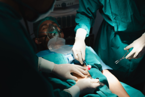
Surgery is done to repair a problem, relieve pain, or improve some body function. Some surgeries are very important, like heart surgery to fix a blocked artery. Other operations can save your life. But many kinds of surgery have risks.
Before surgery, you have tests to make sure you are healthy enough for it. These include blood work, chest x-rays, and an electrocardiogram. Contact Copper Mountain Surgical now!

In this type of surgery, doctors remove a tumor or part of a tumor. They also might recommend chemotherapy or radiation before resection to shrink the tumor, making surgery easier. This can also reduce the chance of tumor recurrence.
In some cases, doctors might need to perform a bowel resection. This is when a cancerous or diseased section of your colon can’t heal correctly. For example, if you have a sigmoid colon tumor, your doctor might need to perform a proctocolectomy and coloanal anastomosis. This will remove your sigmoid colon and connect the healthy ends with an intestinal tube (coloanal anastomosis).
Your doctor might use this procedure to treat other types of tumors in your intestines, stomach, esophagus, or small intestine. Your doctor might perform a biopsy to remove tissue for testing. This procedure is coded to the root operation “excision.” This means that surgeons cut out some or all of a body part without replacing it (partial resection). Some physicians use the terms excision and resection interchangeably. It is important for coders to understand the differences between these two operations so they can accurately assign codes.
Excision
A surgical excision involves the use of a scalpel to cut away damaged or diseased tissue along with some healthy skin around it. It is usually done while the area is numbed with anesthetic. A sample of the tissue is then sent to a lab for testing to make sure the area is free of cancerous cells.
A variety of medical conditions can be treated with surgical excision, including benign tumors and endometriosis. It is a common procedure that can be performed in a hospital or clinic. Before surgery, patients should stop taking any medications that could affect clotting or bleeding. They should also shave the area to be removed and tell their doctor if they have a pacemaker or implanted defibrillator.
Studies show that surgical excision of endometriosis is a safe and effective treatment. However, it is important to choose a surgeon who is experienced in this technique. Your surgeon must know how to recognize the different appearances of endometriosis and have the ability to remove it from many sites in the body.
Ablation
Ablation is a way to treat arrhythmias like atrial fibrillation (AFib) that cause irregular heartbeats and can lead to stroke. This procedure can be used alone or in combination with surgery.
Your healthcare provider puts a thin tube (catheter) into a blood vessel in your groin and threads it up to the heart. A special dye flows through the catheter to help your blood vessels show up better on X-ray images. Sensors on the catheter record your heart’s electrical signals and find where the abnormal ones start.
The healthcare provider uses the catheter to heat or freeze the area of your heart that has abnormal signals. This scars the area and stops it from triggering more arrhythmias.
You can have this procedure under twilight sedation or general anaesthesia. It takes 3 to 6 hours. Complications are rare but can include pain or bleeding where the catheters entered your body, a clot in your lung that needs to be drained, and damage to your normal heart tissue. This can slow your heart rate and lead to a pacemaker being needed later.
Minimally invasive surgery
During minimally invasive surgery, surgeons make very small cuts in your skin — or no cuts at all. Then they insert a scope with a light and tiny video camera on the end. This gives the surgeon a magnified view of the surgical site. Surgeons then use specialized tools to perform the surgery. This type of surgery reduces the risk of complications and allows you to recover faster.
Minimally invasive surgeries are often used for patients who need surgery because of cancer, arthritis, chronic pelvic pain and other conditions. They are also used for people who have a hard time with large incisions. For example, older people who undergo abdominal procedures with large incisions may be at higher risk for blood clots that can cause serious medical problems.
Many of the surgeons at Yale Medicine are leaders in minimally invasive techniques and routinely perform thousands of these procedures, including robot-assisted surgery. Our doctors believe that this type of surgery helps them treat patients more effectively, so they can provide whole-person care for the patient and family.
Incisions
A surgical incision is a cut that surgeons make in the skin to expose tissues and facilitate an operation or procedure. A variety of conditions require surgical treatment, and the type of incision will vary depending on the procedure. Taking proper care of your incision promotes healing and minimizes scarring. It’s important to follow your healthcare provider’s instructions for wound care. If your incision is bleeding excessively, oozing or soaking through dressings, see your doctor immediately.
A thoracic incision is made on the chest (thorax) to access the space between the lungs and chest wall. It’s usually performed for lung cancer, noncancerous lung masses, thoracic aortic aneurysm (ballooning or bulging of the aorta in weak areas of its wall), esophageal tumors and chest injuries. The McBurney incision is a special incision that’s used to perform appendectomy. It’s made at the junction of two-thirds of the abdominal line running from the umbilicus to the anterior superior iliac spine. It’s also common to use Lanz and gridiron incisions during gallbladder removal operations.
Endovascular catheters
A catheter is a hollow tube that can be used to diagnose or treat diseases in blood vessels throughout the body. Interventional radiologists use catheters for diagnostic procedures like angiographies (X-rays of the heart), and for treating coronary artery disease, renal stenosis, abdominal aortic aneurysms, and brain tumors.
To guide catheters to their destinations, interventional radiologist use a technique called fluoroscopic guidance, which requires X-rays and contrast injections. Our team has developed a new approach that uses bioelectric feedback to identify landmarks and track the position of a catheter in blood vessels without imaging or contrast.
To place a PICC line, the physician or a nurse will insert a needle into an arm vein and then advance a guide wire into a large central vein in the chest called the superior vena cava, which is visible on an X-ray (fluoroscopy). Then, the doctor inserts the catheter over the guide wire. The PICC line is tunneled partially or completely beneath the skin and is designed to remain in place for long-term access to chemotherapy drugs.
Trocars
Trocars are surgical instruments that create a passage through which other instruments can be inserted into a body cavity. They are used in laparoscopic surgery, a minimally invasive technique that allows surgeons to perform procedures that would otherwise require making large abdominal incisions. Trocars are a key component of laparoscopic surgery and can help prevent complications like hernias and postoperative pain.
A trocar consists of two parts: the cannula, which is pushed through the skin and subcutaneous tissue to reach a body cavity, and the obturator, which is fitted on top of the cannula and has either a sharp point or a blunt tip. The cannula has a seal at the top to maintain air pressure inside the body cavity.
Trocars may be disposable or reusable. Disposable cutting trocars have a sharp blade and are used to cut through the skin, while reusable trocars have a blunt obturator that is used to push apart the tissue instead of cutting it. The use of a blunt-tip noncutting trocar is associated with a lower risk of vascular and visceral injury than the use of a cutting trocar (Peto OR 0.28, 95% CI 0.014 to 0.54, five studies, 654 participants). However, this finding is not sufficient to demonstrate a benefit for one type of trocar over another effectively.
Insufflators
A surgical insufflator is a device used to inflate body cavities with medical gas such as carbon dioxide. It is useful for laparoscopic examinations and surgeries. It also dilates the peritoneal cavity, allowing the surgeon to have sufficient workspace for their procedure. This is essential for good visibility and precise incisions.
The insufflator keeps intra-abdominal pressure at a preset level and ejects some air when the actual intra-abdominal pressure decreases due to leakage from the port. It also sucks some air from outside when the actual intra-abdominal is higher than the preset pressure to prevent gas embolism.
Insufflators contain several sensors that control the pressure and flow of gas into the abdominal cavity. They require periodic servicing and testing to ensure that the sensors are working properly. These tests are typically conducted by the manufacturer or an authorized service technician. The test results should be recorded in the patient’s medical record. This will help the physician determine whether the insufflator is preventing laparoscopic hypothermia or not. We studied the effectiveness of insufflators with internal gas heating versus those without at different gas flow rates. We found that insufflators with internal gas heating were unable to increase the abdominal gas temperature sufficiently and should not be relied upon for hypothermia prevention.